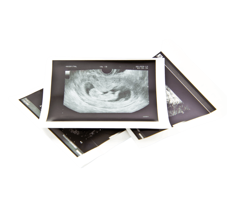
Growth Ultrasound
The third trimester is the phase of pregnancy when everything accelerates. The baby gains weight, movements become stronger, and the due date draws closer.
The growth ultrasound is the examination that tells us whether this course is progressing as it should—whether the baby is growing properly, whether the placenta is functioning well, and whether the amniotic fluid is within normal levels.
What is a growth ultrasound and what does it show?
A growth ultrasound is a systematic ultrasound assessment that evaluates fetal growth and condition in the third trimester.
Unlike the second-trimester anatomy scan, which focuses on anatomy, here the goal is the fetus’s growth, weight, and well-being.
During the examination, we assess:
Fetal biometric measurements
We measure the fetus’s key dimensions (head diameter, abdominal circumference, femur length) and use these to calculate the estimated fetal weight. The weight is then compared with normal values for the corresponding gestational week.
Amniotic fluid
We assess the amount of amniotic fluid; both a reduced amount (oligohydramnios) and an increased amount (polyhydramnios) are findings that require monitoring.
Placenta
We check the placenta’s position, texture, and maturity—features that give us information about its function and the adequacy of fetal nourishment.
Fetal position
In the third trimester, fetal position begins to gain clinical significance, with important implications for the mode of delivery.
Cardiac activity
We confirm normal fetal heart function and rhythm.
When is the growth ultrasound performed?
The growth ultrasound is performed from the 28th week until the end of pregnancy, with the ideal timing being around 30–32 weeks.
It is not an examination performed only once. In high-risk pregnancies or when there are findings that require follow-up, it may be repeated every 2–4 weeks until delivery.
In uncomplicated pregnancies, one or two growth ultrasounds in the third trimester are usually sufficient.

How is it done and how long does it take?
It is performed transabdominally, with the probe on the abdomen, and is completely painless and safe for the fetus. No special preparation or fasting is required. It is recommended that you empty your bladder before the examination so that you feel more comfortable.
The examination usually lasts 20–30 minutes. It is often combined with Doppler flow assessment during the same visit (especially in high-risk pregnancies), so the total duration may be longer.
At the end of the examination, in many cases it is possible to capture the baby’s face in 3D/4D images.
Normal fetal weight — What to expect by week
One of the most common questions after a growth ultrasound is: “Is my baby’s weight normal?”
Indicative values:
- 28 weeks: ~1,000–1,100 g
- 30 weeks: ~1,300–1,500 g
- 32 weeks: ~1,700–1,900 g
- 34 weeks: ~2,100–2,300 g
- 36 weeks: ~2,500–2,700 g
- 38 weeks: ~3,000–3,200 g
- 40 weeks: ~3,300–3,500 g
Note: These are average values—there is normal variation. What matters most is not the absolute number but the growth curve, that is, whether the fetus is growing steadily over the weeks.
What does “small for gestational age” mean?
If the estimated fetal weight or measurements are below the 10th percentile for gestational age, we speak of intrauterine growth restriction (IUGR). This does not automatically mean there is a problem. Some fetuses are constitutionally small, simply reflecting their parents’ genes.
What differentiates a “small but healthy” fetus from one with true growth restriction is the combined assessment: growth curve, umbilical artery Doppler flow, amniotic fluid volume, and the overall clinical picture. That is why every finding is always evaluated as a whole and not in isolation.
What is the biophysical profile?
The biophysical profile is an additional way to assess fetal well-being that is usually used after 32 weeks, mainly in pregnancies where there is a finding that requires monitoring.
During it, we observe and score four parameters:
- Fetal breathing movements — is the baby “practicing” breathing?
- Body and limb movements — how much and how does the baby move?
- Muscle tone — does the baby open and close their hands and feet?
- Amniotic fluid volume — is it adequate?
Assessment in the final weeks (after 35 weeks)
As delivery approaches, the growth ultrasound gains additional clinical importance. In addition to growth and weight, we also assess:
Fetal position (cephalic, breech, or transverse)
This position largely determines the mode of delivery and is one of the most common questions during the final visits.
Umbilical cord position
We check whether the umbilical cord is around the neck or in front of the baby’s head—findings that are taken into account when planning delivery.
Placental maturity and position
We make sure that the placenta is not near or covering the cervix (placenta previa).
Condition of the cesarean scar
In women with a previous cesarean section, we assess the integrity of the scar in view of the upcoming delivery.

Frequently Asked Questions (FAQ)
In an uncomplicated pregnancy, usually one (around 32 weeks). In high-risk pregnancies or when there are findings that require follow-up, the examination is repeated more often.
No, they are two different examinations, although they are often performed together. The growth ultrasound measures the fetus’s dimensions and weight; Doppler assesses blood flow in the vessels. Together, they provide a much more comprehensive picture.
Yes, especially up to 34–35 weeks the fetus has enough room to change position. After 36 weeks, the position gradually stabilizes, although exceptions do occur.
A reduced amount of amniotic fluid is always evaluated together with the other findings and the gestational age. In some cases, closer monitoring or even early completion of the pregnancy may be required. The decision is always made on an individualized basis.
Get a second opinion! Schedule an appointment